Joshua
Place on VV ECMO
Place on VA ECMO
Place on VA-V ECMO
Decline for ECMO Support
Joshua falls into a quagmire where his hypercarbia worsens his ICP, yet so does the impact of high intrathoracic pressures on his CVP, and thereby his ICP. Anticoagulating a patient with traumatic intracranial hemorrhages is a bit frightening at best; but with care, it can be done safely. We will describe his course and then discuss the rationale, and alternative approaches.
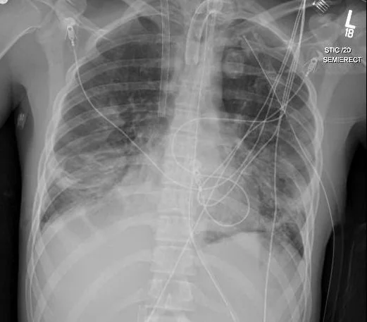
In preparation for cannulation, baseline coagulation studies including an ACT and a TEG with LY30 were checked, and he was then prepped for femoral and IJ cannulation. Once wire access was obtained, he was given a 100U/kg bolus of unfractionated heparin, followed by a 100g/kg bolus of the antifibrinolytic Amicar (epsilon aminocaproic acid), and then the cannulas were inserted and connected to the circuit. Flow was established, the ventilator settings were reduced to “rest” mode to protect his lungs and decrease his CVP, and the circuit sweep gas was used to reduce his pCO2 gradually, with a dramatic drop in his pCO2, likely from both actions. Low level heparin anticoagulation infused into the venous side of the circuit was maintained with lower anti-Xa goals, and an Amicar infusion at 30mg/kg/hr was started after the initial bolus, with periodic TEG-LY30 checks to make sure we were suppressing fibrinolysis sufficiently. On ECMO day 2 Joshua had a repeat head CT (below) and also had a tracheostomy performed at bedside. With his continued improvement in his intracranial pressures, the pentobarbital was stopped, and the ventricular drain was removed. After 13 days of VV support he had a final CXR and a repeat head CT (below) and he was successfully weaned and decannulated from ECMO. He subsequently woke up and was weaned to trach collar 1 week post decannulation. He later left the ICU, and after 2 more weeks he was transferred to rehab, and after three weeks there he was finally able to go home, with continued out-patient therapy and follow-up for his traumatic brain injury.
Some additional points on Joshua’s case management:
Trauma and neurosurgery colleagues expressed a lot of concern about the saturations in the low 80’s on VV ECMO regarding its effect on the injured brain. But the brain actually gets more oxygen delivery with an excellent cardiac output and brain perfusion with a saturation of 80%, than with a saturation of 96% on high vent settings, decreased cardiac output, and poor brain perfusion due to the high ICP and CVP.
This and several other very similar cases with similar positive results may suggest the CT scan finding of “sheer injury” or “diffuse axonal injury” as a predictor of poor neurologic outcome may not be completely accurate when these patients are offered ECMO as a means of reducing ICP and improving brain perfusion and oxygen delivery. The question arises if perhaps, like some other historical predictors of outcome, these findings become a self-fulfilling prophecy if they lead to withdrawal of care. More than one person suggested that ECMO for Joshua was “futile” given his CT scan findings.
Heparin doesn’t cause bleeding. It prevents clotting. Those are not the same thing. Much of the bleeding complications on ECMO are actually due to the activation of fibrinolysis by exposure of the blood to the ECMO circuit, primarily the oxygenator. Once recent clots are lysed, bleeding starts again and then it is more difficult to stop because of the heparin. With low levels of anticoagulation into the venous side of the circuit to prevent circuit thrombosis, with lower anti-Xa goals, and with the use of antifibrinolytics such as Amicar or tranexamic acid (TXA) with TEG-LY30 monitoring (or other methods) patients with recent surgery or injuries that are at least a day or two old, and are not actively bleeding, have been be safely managed on ECMO.
Anticoagulation free ECMO has become popular because of concerns over bleeding risks and frustrations managing anticoagulation, despite a paucity of reports, and a complete absence of any prospective randomized trials. Remember just because you can get away with something doesn’t mean it is a good idea. In patients that are actively bleeding, running ECMO without anticoagulation can be done “safely”, but not without risk. And sudden circuit thrombosis can be lethal. Several steps can be taken to reduce the risk of a clotted circuit, and to be better prepared to handle it when it occurs:
Kepp the flow as high as you can practically to minimize the risk of thrombosis in the circuit.
Closely monitor the transmembrane pressure.
Avoid high volume and/or rapid transfusion of FFP, cryoprecipitate and platelets to the patient (and NEVER to the circuit) when treating a patient’s coagulopathy.
Have a primed circuit, or a primed oxygenator close at hand that can be quickly spliced in to rescue the patient (including the clamps, scissors, etc).
For patients who fail to wean from CPB and are transitioned over to VA ECMO, consider the use of a Carmeda (heparin bonded) coated oxygenator such as the Medtronic Affinity NT or Fusions polypropylene membranes. These can be safely used for up to 48 hours without heparin, with close monitoring. They can also be inserted for non-cardiac surgery patients who are having significant bleeding at the time of ECMO initiation, such as trauma patients.